Scanning Eletron Microscopy photograph of the leaf surface of Solanum
By A Mystery Man Writer
Description
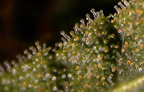
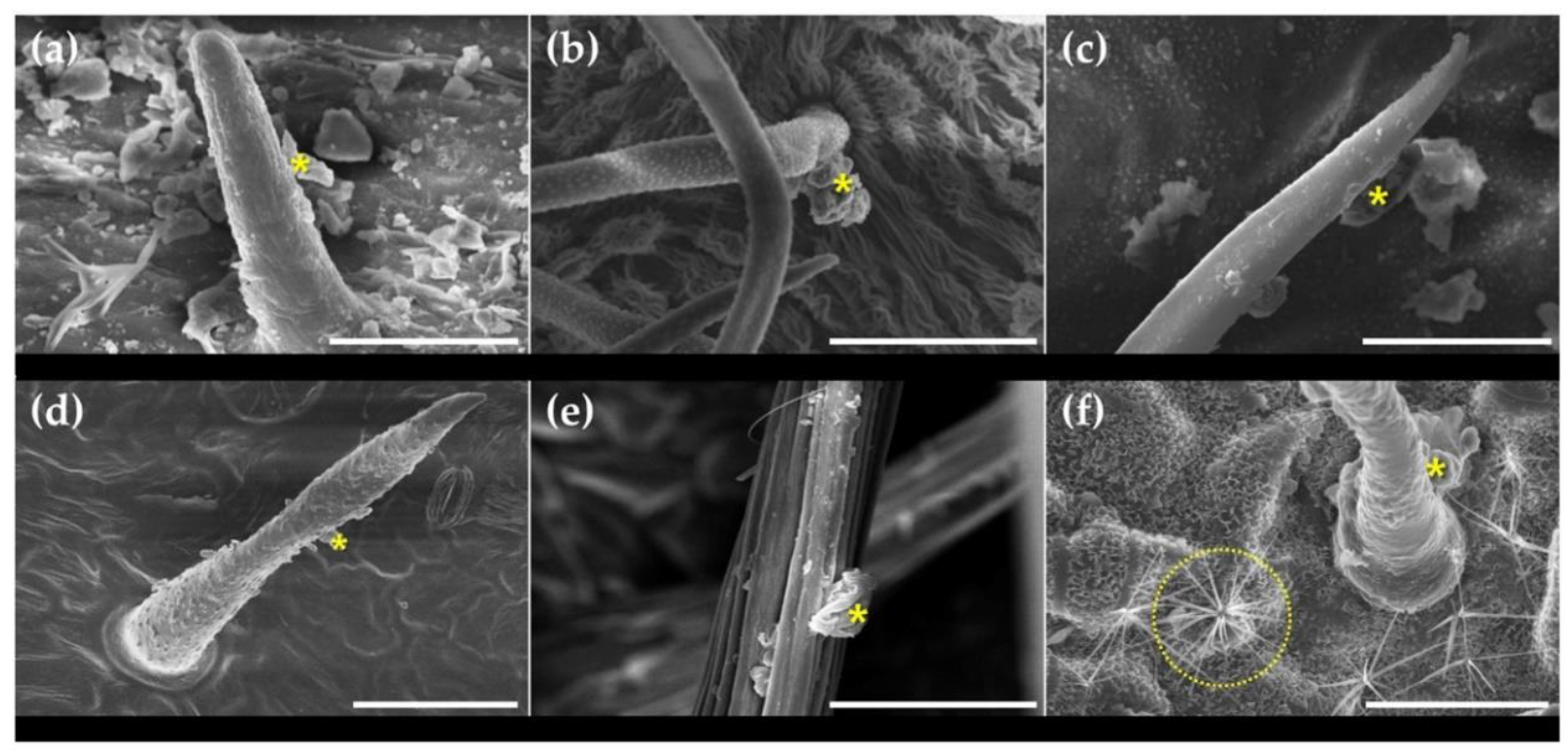
Download scientific diagram | Scanning Eletron Microscopy photograph of the leaf surface of Solanum granuloso-leprosum Dunal. A – Unicelular tector trichomes; B – tector trichome, note that there are projections at the trichome base; C – tector trichome, note that there is a larger projection/ramification at the trichome base; D – tector trichome, note that there are two larger projection/ramification at the trichome base; E – tector trichome, note that there are three larger projection/ramification at the trichome base; F – tector trichome, note that there are four larger projection/ramification at the trichome base; G – tector trichome, note that there are five larger projection/ramification at the trichome base; H – tector trichome, note that there are six larger projection/ramification at the trichome base; I – tector trichome, note that there are eight larger projection/ramification at the trichome base; J – another angle from the six ramification tector trichome; and K – multicelular and multisseriated tector trichome, note the thick secondary cell wall. Scale Bars = 20 μm. from publication: Anatomy, histochemistry and micromorphology of leaves of Solanum granuloso-leprosum Dunal | In the present work the anatomical, histochemical and micromorphological features of S. granuloso-leprosum leaves were approached in order to evaluate its characteristics associated with its pioneer role. Glandular and non-glandular trichomes were observed on both epidermal | Micromorphology, Solanum and Plant Anatomy | ResearchGate, the professional network for scientists.

File:Leaf epidermis.jpg - Wikipedia
Scanning electron microscope image plant hi-res stock photography and images - Alamy

Herbs Micronaut: The fine art of microscopy by science photographer Martin Oeggerli
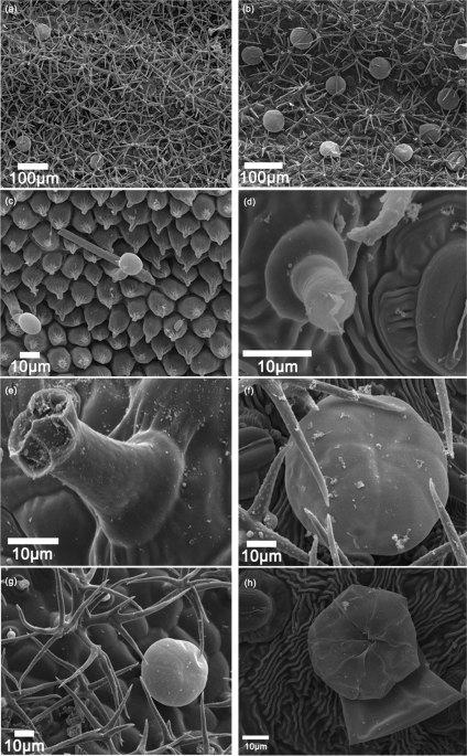
Scanning electron microscopy (SEM) micrographs of the adaxial leaf
Forests, Free Full-Text
Accumulation and transfer of polystyrene microplastics in Solanum nigrum seedlings [PeerJ]
Image library - substance type - Quorum Technologies Ltd
Insects, Free Full-Text
Formation mechanism of glandular trichomes involved in the synthesis and storage of terpenoids in lavender, BMC Plant Biology
from
per adult (price varies by group size)